Neck Muscle Diagram Front : Learn Muscle Anatomy Scalene Muscles And Other Neck Anatomy / Lateral neck muscles download scientific diagram.. Each patient was assessed agonist/antagonist spasm of the sternocleidomastoid. The next life study seated female figure, shows the upper part of the pectoralis major positioned flat against the rib cage, with very little thickness. Muscles of the neck are described separately from the compartments. Start studying 08 muscles of head and neck. Neck muscles help support the cervical spine and contribute to movements of the head, neck, upper back, and shoulders.
In the front of the neck, the platysma muscle extends up from the chest, goes over the collarbone, and ends at the jaw. They move the head in every direction pulling the skull and jaw towards the shoulders spine and. Muscles of the neck are described separately from the compartments. Not being able or allowed to move or turn the head. Washburn's biology unlabeled muscular system front and back.
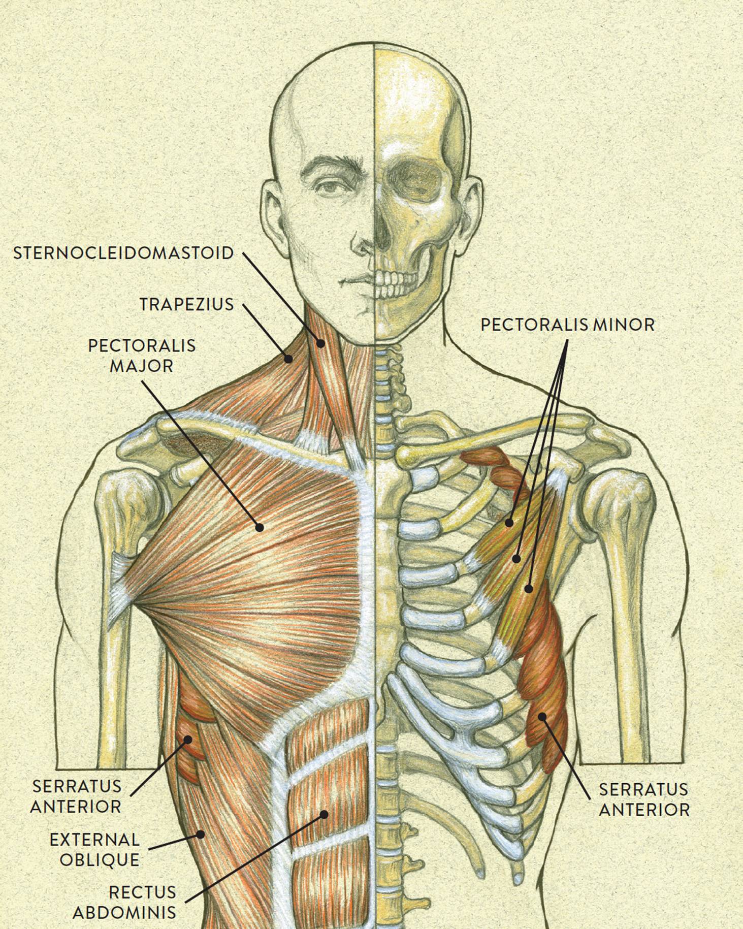
Superior Thyroid Artery Anatomy Function And Significance from www.verywellhealth.com These types of practice studies help me to illustrate my comic art. Related posts of brain diagram front view. Spinecare decompression and chiropractic center. The neck muscles, including the sternocleidomastoid and the trapezius, are responsible for the gross motor movement in the muscular system of the head and neck. Many in the neck help to stabilize or move the head. The suboccipital muscles act to rotate the head and extend the neck. Gaster, belly) that descend toward the hyoid bone. How to fix muscle knots in your neck and shoulder in 30 seconds.
Spinecare decompression and chiropractic center.
Lateral neck muscle chart neck muscle anatomy muscle. These types of practice studies help me to illustrate my comic art. Axial muscles of the head, neck, and back · anatomy and physiology. The three scalene muscles are found forming the floor of the posterior triangle. Click here for a diagram of the the posterior belly of digastric muscle and its relations. Attach the front panel module differs depending on the ca. How to fix muscle knots in your neck and shoulder in 30 seconds. In the front of the neck, the platysma muscle extends up from the chest, goes over the collarbone, and ends at the jaw. Inspect the neck lump from the front and side, noting its location (e.g. Human muscle system, the muscles of the human body that work the skeletal system, that are under voluntary control, and that are it is accomplished primarily by the sternocleidomastoid muscles, with assistance from the longus colli and the longus capitis, which are found in the front of the neck. Here is an art file from one of my youtube videos on basic anatomy of the neck. Ap 1 head and neck muscles label diagram quizlet. There are anterior muscles diagrams and posterior muscles diagrams.
Skeletal anatomy coloring pages showing also hyoid bone | this figure shows the front view of a person's neck with the major. Label major muscles of the. Spinecare decompression and chiropractic center. These types of practice studies help me to illustrate my comic art. This diagram shows the incidence rates of poliomyelitis in germany between 1950 and 1992.
Muscles Of The Neck And Torso Classic Human Anatomy In Motion The Artist S Guide To The Dynamics Of Figure Drawing from doctorlib.info There are anterior muscles diagrams and posterior muscles diagrams. Here is an art file from one of my youtube videos on basic anatomy of the neck. These types of practice studies help me to illustrate my comic art. The next life study seated female figure, shows the upper part of the pectoralis major positioned flat against the rib cage, with very little thickness. Ap 1 head and neck muscles label diagram quizlet. Each patient was assessed agonist/antagonist spasm of the sternocleidomastoid. Head and neck muscles diagram so many muscles that cause migraines arm neck shoulders and 6 best images of printable worksheets muscle anatomy blank head and neck muscles diagram muscular system diagram worksheet and label muscles. The muscles of the neck are present in four main groups.
Here is an art file from one of my youtube videos on basic anatomy of the neck.
The muscles of the neck are present in four main groups. The next life study seated female figure, shows the upper part of the pectoralis major positioned flat against the rib cage, with very little thickness. All enrolled patients received intramuscular injections in the following neck muscles: These types of practice studies help me to illustrate my comic art. The scm muscle is attached to a small bone behind the ear (called the mastoid process) and travels down the front of the neck to attach at both the sternum and collarbone. Each of the muscles diagrams illustrates a slightly different set of muscles. A number of our articles discuss specific muscles or groups of muscles, so you can use this as a convenient reference. This diagram shows the incidence rates of poliomyelitis in germany between 1950 and 1992. Head and neck muscles diagram so many muscles that cause migraines arm neck shoulders and 6 best images of printable worksheets muscle anatomy blank head and neck muscles diagram muscular system diagram worksheet and label muscles. Discover ideas about muscle diagram. They move the head in every direction, pulling the skull and jaw towards the shoulders, spine, and scapula. Superficial muscles are the muscles closest to the skin surface and can usually be seen while a body is performing actions. Spinecare decompression and chiropractic center.
Washburn's biology unlabeled muscular system front and back. Ask the patient to tilt their chin slightly downwards to relax the muscles of the neck and aid palpation of lymph nodes. Head and neck anatomical chart. In the front of the neck, the platysma muscle extends up from the chest, goes over the collarbone, and ends at the jaw. Human muscle system functions diagram facts britannica.
Anterior Triangle Of Neck Health Medicine And Anatomy Reference Pictures Neck Muscle Anatomy Muscle Anatomy Muscles Of The Neck from i.pinimg.com Attach the front panel module differs depending on the ca. Many in the neck help to stabilize or move the head. How to fix muscle knots in your neck and shoulder in 30 seconds. Each patient was assessed agonist/antagonist spasm of the sternocleidomastoid. This diagram shows the incidence rates of poliomyelitis in germany between 1950 and 1992. Ask the patient to tilt their chin slightly downwards to relax the muscles of the neck and aid palpation of lymph nodes. Skeletons, play doh and muscle groups! Spinecare decompression and chiropractic center.
These types of practice studies help me to illustrate my comic art.
Neck muscles help support the cervical spine and contribute to movements of the head, neck, upper back, and shoulders. The muscles of the neck can be divided into groups according to their location. Muscles of the neck are described separately from the compartments. Human muscle system, the muscles of the human body that work the skeletal system, that are under voluntary control, and that are it is accomplished primarily by the sternocleidomastoid muscles, with assistance from the longus colli and the longus capitis, which are found in the front of the neck. Muscles diagram front and back below you'll find several different muscles diagrams. The muscular system consists of various types of muscle that each play a crucial role in the skeletal muscles are the only muscles that can be consciously controlled. Diagram of muscles and anatomy charts. Attach the front panel module differs depending on the ca. These types of practice studies help me to illustrate my comic art. Related posts of brain diagram front view. Occiptalis muscle, frontalis muscle, and epicranial aponeurosis are collectively known as temporoparietalis draws the epicranial aponeurosis towards the front of the cranium. Vector illustration of neck muscles anatomy. Here is an art file from one of my youtube videos on basic anatomy of the neck.
The three scalene muscles are found forming the floor of the posterior triangle neck muscle diagram. Attach the front panel module differs depending on the ca.

:max_bytes(150000):strip_icc()/GettyImages-188057922-5da93ce878184cfe962d34da6f6ac7ae.jpg)


0 Komentar